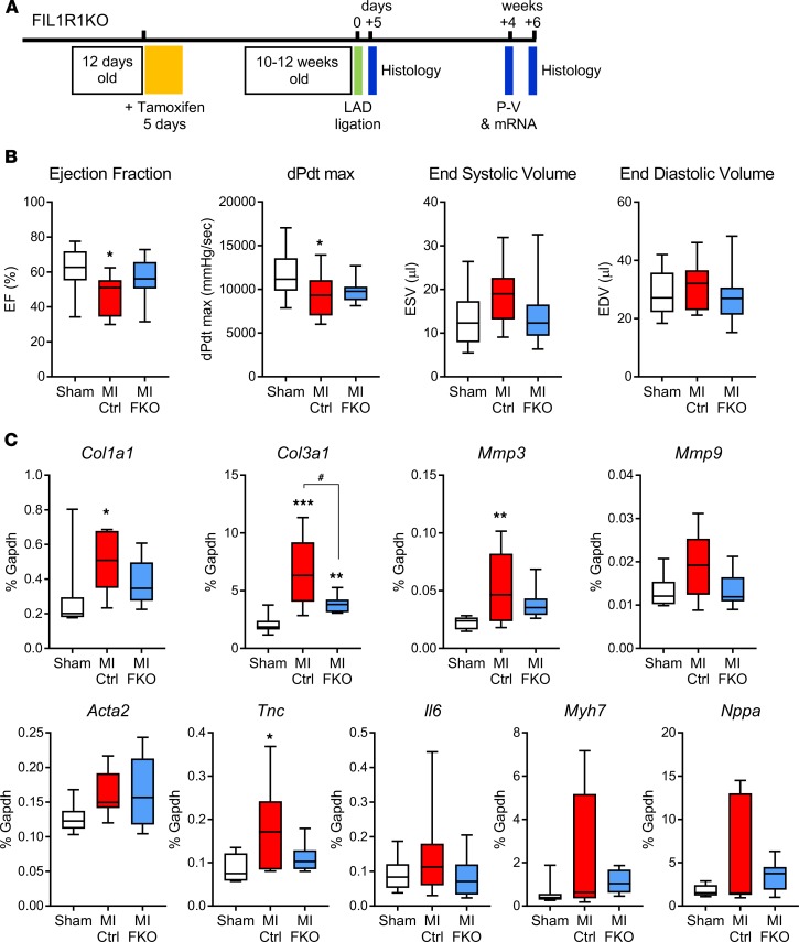
Figure 5. Effect of fibroblast-specific IL-1R1 deletion on cardiac function and remodeling genes 4 weeks after MI.
(A) Experimental timeline showing timing of tamoxifen injection and experimental myocardial infarction (MI) induced by ligation of the left anterior descending (LAD) coronary artery, as well as the timing for histology, collection of RNA, and measurement of cardiac function by pressure-volume (P-V) conductance catheter. (B) P-V conductance catheter data. Sham, mixed genotypes, no tamoxifen treatment (n = 18); MI Ctrl, tamoxifen-treated Cre-negative Il1r1fl/– after MI (n = 11); MI FKO, tamoxifen-treated Cre-positive Il1r1fl/– (FIL1R1KO; fibroblast-specific IL-1 receptor 1 KO) after MI (n = 11). *P < 0.05 versus sham. (C) qRT-PCR data showing relative mRNA levels of remodeling genes collagen Iα1 (Col1a1), collagen IIIα1 (Col3a1), Mmp3, Mmp9, α-smooth muscle actin (Acta2), tenascin C (Tnc), Il6, β-myosin heavy chain (Myh7), and atrial natriuretic factor (Nppa). Sham, mixed genotypes, no tamoxifen treatment (n = 8); MI Ctrl, tamoxifen-treated Cre-negative mice after MI (n = 8); MI FKO, tamoxifen-treated FIL1R1KO mice after MI (n = 8). *P < 0.05; **P < 0.01; ***P < 0.001 versus sham; #P < 0.05 versus MI Ctrl (1-way ANOVA with Tukey post hoc).

