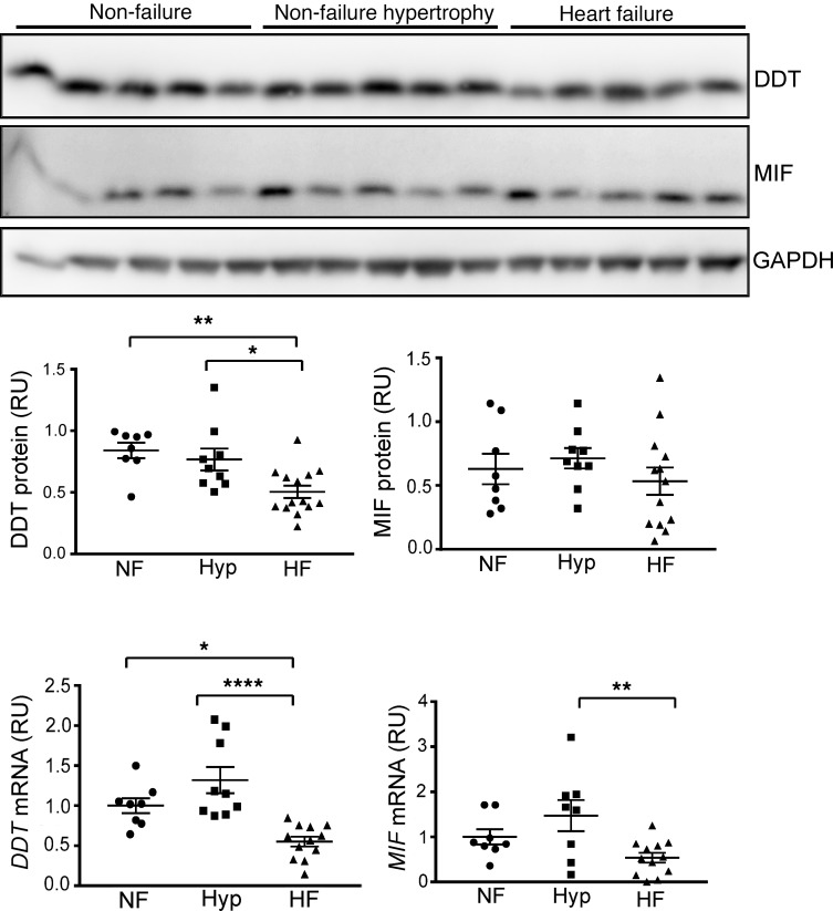
Figure 1. Cardiac DDT and MIF expression in human heart failure.
LV myocardial tissue excised from nontransplanted donor nonfailing hearts (with or without hypertrophy) or from explanted hearts from patients undergoing cardiac transplantation for heart failure due to ischemic or nonischemic cardiomyopathy. Upper, immunoblots showing human heart tissue homogenate DDT, MIF, and GAPDH content in nonfailing, nonfailing hypertrophy, and cardiomyopathy hearts. Lower, human cardiac DDT and MIF protein and mRNA content quantified relative to GAPDH. NF, nonfailure; Hyp, nonfailure hypertrophy; HF, heart failure; RU, relative unit. Data are shown as mean ± SEM; n = 8 for the nonfailing heart and nonfailing hypertrophy heart; n = 12 for the failing heart group. Significance determined by 1-way ANOVA with Tukey’s multiple-comparisons test. *P < 0.05; **P < 0.01; ****P < 0.0001 indicated by brackets.

