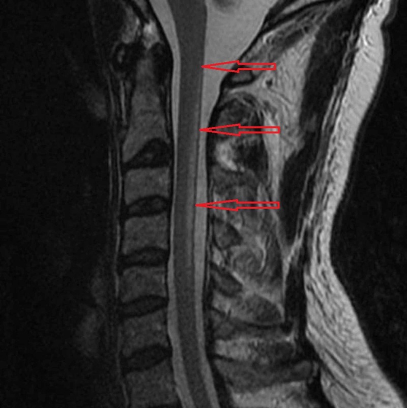
Figure 1. MRI reveals abnormal high T2 cervical cord signal within the dorsal columns extending from C2 to C5 highlighted by the red arrows.
The location of the signal abnormality is consistent with subacute combined degeneration of the cord.
Image courtesy: [11]

