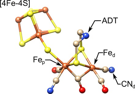
Fig. 2. The molecular structure of the [FeFe]-hydrogenase active site, the H-cluster.

Highlighted are the proximal and distal irons, Fep and Fed, respectively, the cyanide ligand (), and the ADT ligand. S, yellow; Fe, orange; N, blue; C, tan; O, red. Structure is from Protein Data Bank (PDB) ID 4XDC.
