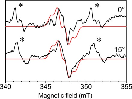
Fig. 4. Single-crystal continuous-wave EPR of in the photosystem II core complex.

Continuous-wave EPR collected with the 0.4 mm inner diameter self-resonant microhelix at two angles of the photosystem II radical from a single crystal at a temperature of 80 K. The crystal dimensions were 0.3 mm by 0.18 mm by 0.18 mm. Shown in red is a fitted simulation with similar features. A nonspecifically bound Mn2+ signal is also present in the mother liquor of the crystal, indicated by an asterisk (∗). Each spectrum was collected in 49 min with a signal-to-noise ratio of approximately 35.
