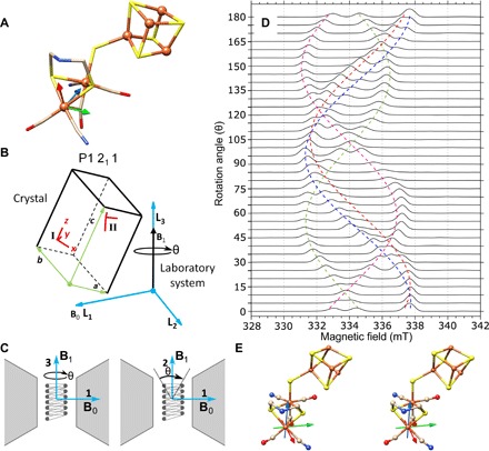
Fig. 5. Pulse EPR on a single crystal of the H-cluster in [FeFe]-hydrogenase.

(A) The molecular structure of the [FeFe]-hydrogenase active site, the H-cluster, from PDB ID 4XDC is shown with the molecular frame located with the distal iron (Fed) as the origin. The molecular frame rotational coordinates can be found in table S2. S, yellow; Fe, orange; N, blue; C, tan; O, red. (B) The P1211 symmetry schematic relating the molecular frame (x, y, z) to the crystal frame (a, b, c) and, last, to the laboratory system frame (L1, L2, L3) is shown. The two molecular frames from the asymmetric unit are present in Site I and can be translated to Site II by crystal symmetry operations. (C) The static magnetic field (B0) is positioned along the L1 axis, while the microwave magnetic field (B1) can be either along the L2 axis or along the L3 axis. A rotation of 180° is feasible around the L3 axis, but only a partial rotation around the L2 axis is feasible because of the B1 rotating with the crystal resulting in B1 to become parallel to B0. A third partial rotation is feasible if the sample is rotated by 90° around the L2 axis. (D) Pulse EPR experiments collected with the 0.4 mm inner diameter self-resonant microhelix with a [FeFe]-hydrogenase single crystal of C. pasteurianum (CpI) in the Hox state showing collected data in one plane for a full rotation of 180° in 5° steps at a temperature of 15 K. The crystal dimensions were approximately 0.3 mm by 0.1 mm by 0.1 mm, and each spectrum was collected in 8 min with a signal-to-noise ratio of approximately 290. (E) A stereo view of the analyzed g-tensor (gx, red; gy, green; and gz, blue) is mapped on the crystal structure (PDB ID: 4XDC). For a three-dimensional (3D) view of the proposed g-tensor, see https://act-epr.org/FeFeHydrogenase.html.
