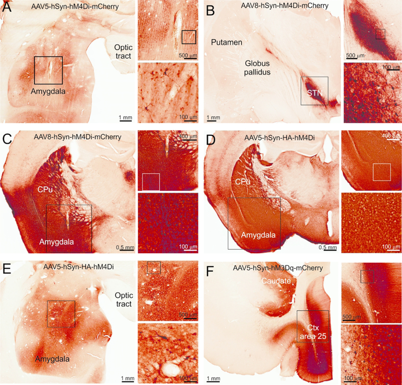
Figure. 1: Representative images showing expression of DREADDs in monkey and mouse brain tissue after viral vector injections.

The immunoperoxidase method was used to reveal the tag proteins fused to the DREADDs. All the sections are adjacent or at the site of the injection track. A) Monkey MR264, B) Monkey MR275, C) Mouse RM9, D) Mouse RM34, E) Monkey MR290, F) Monkey MR293. In each panel, the rectangles indicate areas that are shown at higher magnification in the subsequent images.
