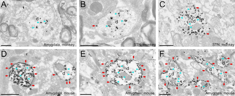
Figure 2. Ultrastructural localization of hM4Di-mCherry in monkeys and mice.

The pre-embedding immunogold method was used to reveal the hM4Di-mCherry. A to C: Electron micrographs showing examples of labeled dendrites in monkey amygdala (A) or STN (B, C). Immunogold particles labeling for hM4Di-mCherry is found mostly in the intracellular compartment (blue arrowheads). D to F: Electron micrographs of labeled dendrites in mice amygdala. The bulk of hM4Di-mCherry gold particles labeling is bound to the plasma membrane (red arrows). All scale bars= 0.6 μm. d, dendrite.
