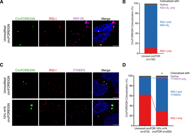
Figure 6. Immunofluorescence reveals circFOREIGN co-localization with RIG-I and K63-Ubn and YTHDF2 recruitment to m6A-modified circFOREIGN.
A. CircFOREIGN co-localizes with RIG-I and K63-polyubiquitin chain. Representative field of view is shown.
B. Quantification of circFOREIGN colocalization with RIG-I and K63-Ubn (n = 152). Foci were collected across 10 fields of view across biological replicates and representative of replicate experiments.
C. 10% m6A circFOREIGN has increased co-localization with YTHDF2. Representative field of view is shown. Foci were collected across >10 fields of view and representative of replicate experiments.
D. Quantification of circFOREIGN and 10% m6A circFOREIGN colocalization with YTHDF2 and RIG-I. *p<0.05, Pearson’s χ² test.

