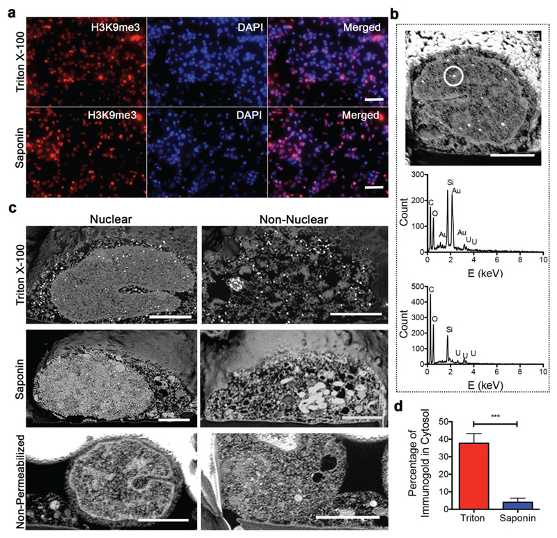
Figure 2. Optimization of immunogold FIB-SEM.
a) Widefield fluorescence images of stem cells stained with DAPI, H3K9me3 primary antibody, and FluoroNanoGold secondary antibody after permeabilization with either Triton X-100 or Saponin. Scale bars = 20 μm. b) FIB-SEM cross section of a neural stem cell nucleus immunolabeled and prepared according to the workflow (top), with corresponding EDX spectra of the circled region indicating the presence of gold (middle). EDX spectra of a negative control with no H3K9me3 primary antibody added showed no gold present (bottom). Scale bars = 2 μm. c) Nuclear and non-nuclear FIB-SEM cross sections of neural stem cells either permeabilized with Triton X-100 or Saponin or not permeabilized at all. Scale bars = 2 μm. d) Quantification of immunogold particles for nuclear antigen H3K9me3 in the cytosol of samples permeabilized with Triton X-100 and Saponin as a percentage of the total visible labels. Plot shows mean ± standard deviation (S.D.), n = 3 (cells), *** p < 0.001 (two-tailed Mann–Whitney test).

