Fig. 1.
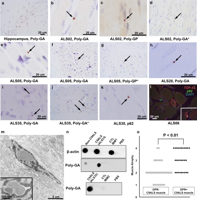
DPR pathology in postmortem C9ALS muscle samples. a Poly-GA staining in the dentate gyrus of a C9ALS patient. b-j Poly-GA and poly-GP immunohistochemistry reveals discrete, perinuclear DPR inclusions (black arrows) in muscle samples of two patients from MCJ (b–g) and two from HMH (h–j). Staining shown performed at both HMH and MCJ (latter examples are indicated by asterisks). k p62 perinuclear inclusion in case ALS35, which also had conspicuous poly-GA and poly-GP inclusions. l TDP-43 and p62-positive foci in a C9ALS muscle (inset shows an additional focus in the same patient), shown here by double-labeling immunofluorescence. Supplemental Fig. 1 shows separate color channels in two samples. m Electron microscopy of a DPR-positive sample showed nuclei with homogeneous, granular intranuclear material as well as tubule-like structures in close proximity to subsarcolemmal nuclei (inset). n Dot blot (top) of ALS without C9ORF72 expansion (labeled “Non-C9ALS”) (left), C9ALS (middle), and control/ IBM muscle (right) showed poly-GA in C9ALS but not other samples. Repeat dot blot (bottom) in C9ALS versus an additional control sample showed the same. o DPR-positive C9ALS muscle samples were associated with significantly greater muscle atrophy than DPR-negative samples. For a–l all images were photographed with a 60 × objective. The scale bar in a applies to d, f, g, and j, which are not enlarged. b, c, e, h, i, k, and l are enlarged for clarity and each has an image appropriate scale bar

