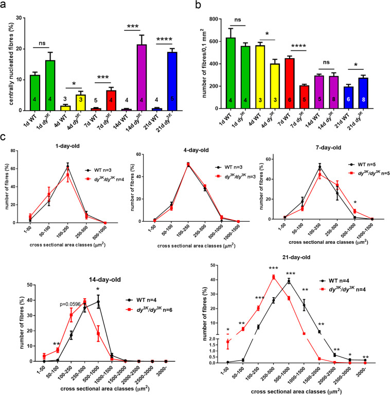
Figure 4.
Morphometric analyses of wild-type and dy3K/dy3K rectus femoris throughout the disease course. (a) Quantification of regenerating fibres. The number of centrally nucleated regenerating myofibres is increased in dy3K/dy3K muscle compared to wild-type muscle from postnatal day 4 onwards (p = 0.0401, p = 0.0004, p = 0.0004, p < 0.0001, for day 4, 7, 14 and 21, respectively). (b) The number of muscle fibres per 0.1 mm2 (rectus femoris). Muscle fibres are temporarily lost at the age of 4 and 7 days in dy3K/dy3K rectus femoris (p = 0.002 and p = 0.0024, respectively). (c) Analysis of muscle fibre sizes from wild-type and dy3K/dy3K rectus femoris (day 1, 4, 7, 14 and 21). Muscle atrophy becomes evident in 14-day-old dystrophic muscles, and the shift towards significantly smaller muscle fibres is particularly pronounced at day 21.

