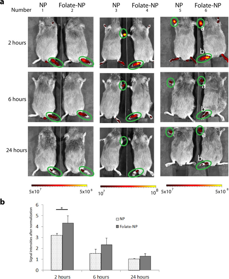
Figure 4.
Imaging of RA in mice by NIR detection. (a) The near infrared images of the dorsal side of arthritic mice at 2, 6 and 24 hours after an intravenous injection of NP or Folate-NP. (b) The quantification of fluorescence signal after normalization within the inflamed foot shown in panel A (green circles). *P < 0.05. Number 6 has two inflamed paws: front paw No. 6a and hind paw No. 6b (Ventral side is presented in supplemental figure 5).

