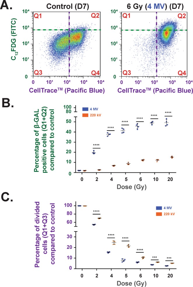
Figure 4.

Cell Division (CellTrace Technology)/ Senescence (C12FDG) (Flow Cytometry) bi-parametric analysis (A) Example of flow cytometry measurements obtained at D7 for control and 6 Gy irradiation at 4 MV. (B) Percentage of β-GAL-positive cells (Q1 + Q2) compared to control at 4 MV (blue dots) and 220 kV (orange dots) irradiations. Each dot represents one independent experiment (****p < 0.001, one-way Anova). (C) Percentage of divided cells (Q1 + Q3) compared to control for 220 kV (orange dots) and 4 MV (blue dots). Each dot represents one independent experiment (***p < 0.001, ****p < 0.001, one-way Anova).
