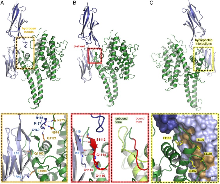
Fig. 2.
The structural basis for ICAM-1 binding by group B PfEMP1. Front views of the DBLβ domain of IT4var13 (green) bound to ICAM-1D1D2 (D1 light blue, D2 dark blue). Dashed boxes highlight the sites that contact ICAM-1 through (A) side chain-mediated hydrogen bonds or (B) a β-sheet augmentation. A third dashed box compares a region of the ICAM-1–bound and unbound conformations of the IT4var13 DBLβ domain. (C) Back view of the IT4var13 DBLβ–ICAM-1D1D2 complex. The dashed box highlights the site of hydrophobic contacts between the DBLβ domain and ICAM-1D1D2 and the residues in the DBLβ domain that contact a hydrophobic patch on the surface of ICAM-1D1 (dark yellow).

