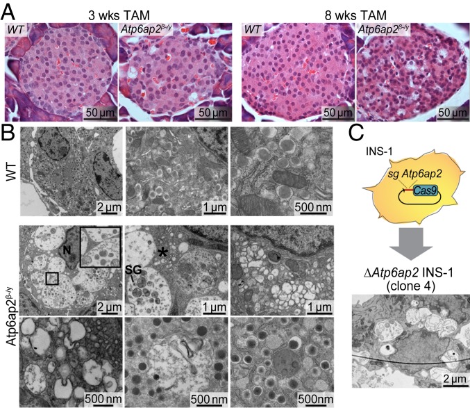
Fig. 1.
Atp6ap2 β cell deletion induces the accumulation of large vacuolated structures. (A) Morphological analysis by hematoxylin and eosin staining in pancreases from WT and Atp6ap2β-/y mice after TAM treatment for 3 and 8 wk. (B) Representative transmission electron microscopy images of β cells from WT and Atp6ap2β-/y mice after 3 wk of TAM. Inset shows a representative single-membraned vacuole in Atp6ap2β-/y islets. N, nucleus; SG, insulin secretory granule; *, dilated Golgi cisternae. (C) Rat insulinoma INS-1 cells were transfected with plasmid expressing Cas9-GFP and sgRNA targeting Atp6ap2. Successful clones deficient for Atp6ap2 (ΔAtp6ap2 INS-1 cells) were analyzed by transmission electron microscopy. Scale bars are indicated.

