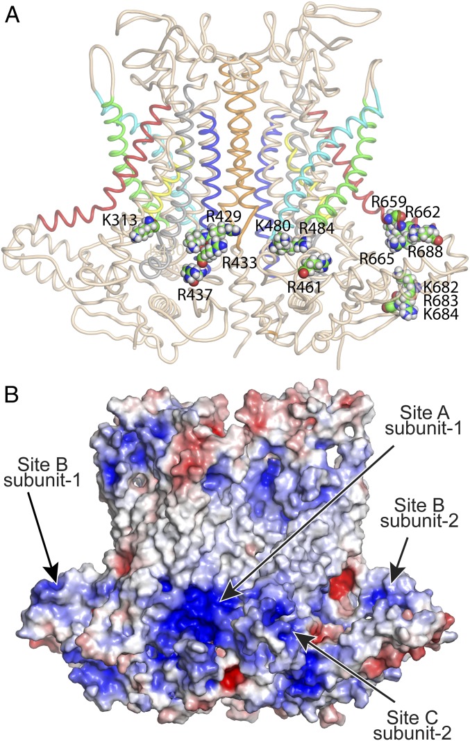
Fig. 4.
Location of mutations that are critical for the stimulatory effect of PI(4,5)P2 on ANO1. (A) Cartoon representation of ANO1 with critical amino acids as space-fill labeled. For clarity, site A (K313, R429, K430, R433, and R437) is shown only in the left subunit; site B (K659, R662, R665, R668, R682, R683, and K684) and site C (R461, K480, and R484) are shown only in the right subunit. Helices are colored: blue (TM2), cyan (TM3), green (TM4), red (TM6), yellow (TM7), and orange (TM10). (B) Electrostatic surface of ANO1 calculated by APBS Electrostatics in PyMOL. Red (+5 kT/e); blue (−5 kT/e).

