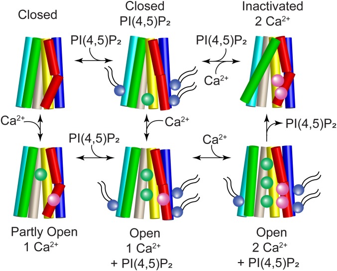
Fig. 9.
Cartoon model of ANO1 gating. TM2 (blue), TM3 (cyan), TM4 (green), TM5 (wheat), TM6 (red), and TM7 (yellow) are shown as cylinders. The pore is formed by TM4–TM7 and Ca2+ (magenta spheres) binds to residues in TM6 and TM7. When PI(4,5)P2 (purple sphere with tails) binds to the cytoplasmic ends of TM2 (site A/1), TM6 (site B/2), and TM3 (site C/4), the cytoplasmic end of TM6 swings away from the pore to ultimately open the cytoplasmic vestibule to Cl− (green spheres). Top row: ANO1 is closed in the absence of Ca2+ and inactivated when 2 Ca2+ ions are bound without PI(4,5)P2. Bottom row: the channel partly opens when one Ca2+ binds without PI(4,5)P2 but full channel opening requires both Ca2+ and PI(4,5)P2.

