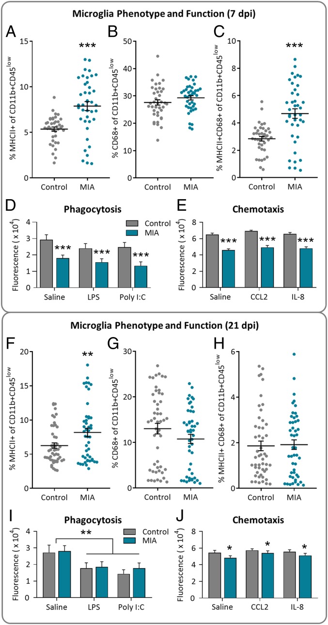
Fig. 2.
Fetal microglia phenotype and function was altered by maternal infection. Maternal infection resulted in an increase in MHCII+ microglia and a reduction in microglia phagocytosis and chemotaxis. (A–E) 7 dpi and (F–J) 21 dpi. (A–E) Percentage of primary CD11b+CD45low microglia expressing (A) MHCII, (B) CD68, or (C) both MHCII and CD68; n = 4 to 5 litters per group, n = 39 to 40 fetuses per group. MIA primary fetal microglia had (D) decreased phagocytic activity and (E) decreased chemotactic activity at 7 dpi. There was no effect of in vitro treatment, n = 7 to 16 fetuses per group; main effect of MIA, ***P < 0.0001. (F–J) Percentage of primary microglia expressing (F) MHCII (main effect MIA, **P < 0.01), (G) CD68, or (H) both MHCII and CD68. n = 5 litters per group; n = 47 to 50 fetuses per group. MIA primary fetal microglia displayed phagocytic activity comparable to controls (I; main effect of in vitro treatment, **P < 0.01) but presented with reduced chemotactic activity (J; main effect of MIA, *P < 0.05; no effect of in vitro treatment) at 21 dpi. n = 7 to 16 fetuses per group; error bars are ±SEM.

