Fig. 4.
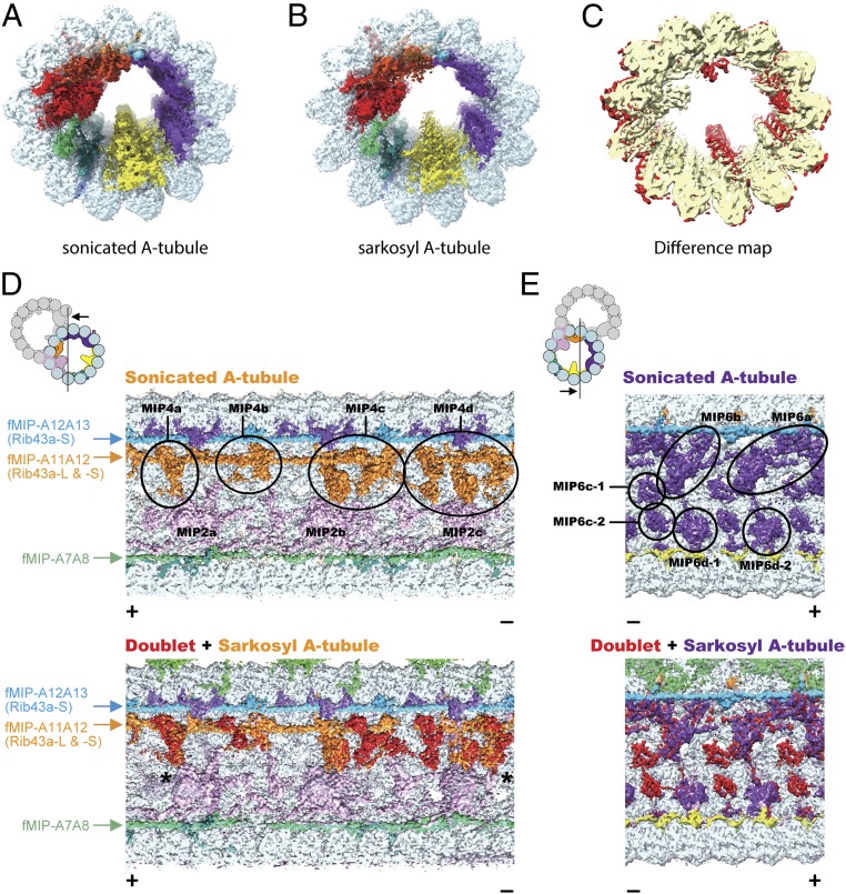
Sarkosyl treatment removes some MIPs from the doublet. (A and B) Surface renderings of the sonicated A-tubule (A) and sarkosyl A-tubule (B) maps. (C) Difference map between the sonicated and sarkosyl A-tubule maps. Superimposition of the 2 maps reveals the missing MIP densities in the sarkosyl A-tubule map (red regions). Parts of the MIP2 and MIP6 are missing in the sarkosyl A-tubule map. (D and E) Sonicated A-tubule map (Top) and the overlap of doublet and sarkosyl A-tubule maps (Bottom). The MIP4 and MIP6 regions of the doublet (red) are mapped onto corresponding regions from the sarkosyl A-tubule map (MIP4 in orange and MIP6 in purple). The views are indicated in the illustrations on the Top Left. Remaining fMIPs are indicated on the side. The coloring of MIP2 and MIP4 is different from other figures to avoid confusion (see the illustration for the coloring). Some densities at the MIP4 and MIP6 regions are missing after the sarkosyl treatment while the fMIPs appear intact. The slight shifts in MIP4 (indicated by asterisks) at both + and − end are due to lateral compaction of the tubulin lattice.

