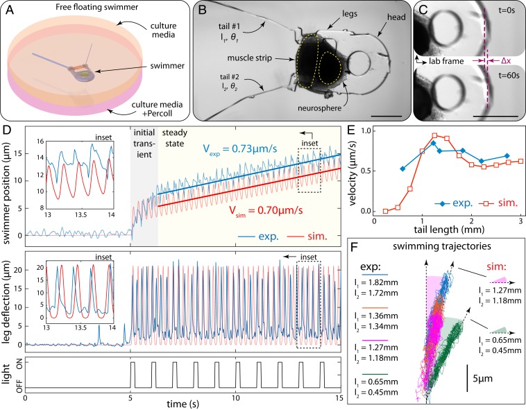
Fig. 4.
Free swimming driven by neuromuscular units. (A) Illustration of free-floating swimmer suspended by Percoll–culture media mixture. (B) Bright-field image of the swimmer after release from the anchors. The neurosphere is dislocated from its original seeding location due to tension generated between muscle and neurons. (C) Forward locomotion of the swimmer illustrated by comparison of snapshots at the beginning (Top) and end (Bottom) of a 60-s recording. (D) Experimental results and model predictions: (Top) swimmer position vs. time, (Middle) leg deflection due to muscle contraction, and (Bottom) input optical signal. (E) Experimental swimming velocity for different average tail lengths compared to model predictions. (F) Swimming trajectories for different tail lengths. (Scale bars in B and C: 500 µm.)

