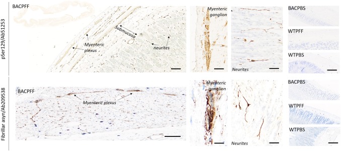
Fig. 5.
Distribution of asyn pathology in the stomach of S129A PFF- and PBS-injected BAC rats and WT controls, at 4 months post-injection, detected with two different antibodies. Scale bar = 100 µm. Representative high magnification photomicrographs are shown of the myenteric ganglion and neurites in the BACPFF. The plexus and lumen of the BACPBS and WT rats remained pathology free. Scale bar: 50 µm

