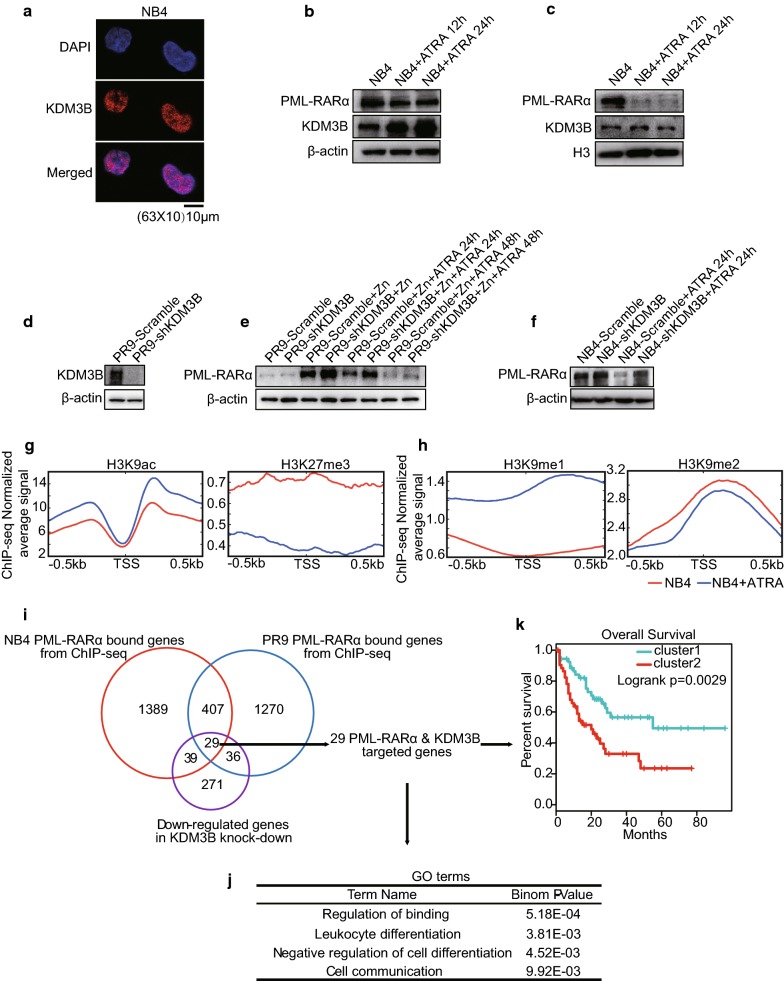
Fig. 5.
KDM3B is associated with PML/RARα oncoprotein during NB4 cell differentiation. a Confocal staining of KDM3B in NB4 cells. Nuclei were counterstained with DAPI. Representative images are shown. Scale bar: 10 μm. b, c Western blot analyses of KDM3B and PML/RARα in NB4 cells at total/nuclear protein level, followed by treatment with ATRA. β-actin was used as a loading control for total protein (b), histone H3 was used for nuclear proteins (c). d Effects of stable KDM3B-knockdown in PR9 cells by Western blot analysis. e Western blot analyses of PML/RARα in PR9 cells transfected with scramble control or shKDM3B, with or without Zn2+ pretreatment for 4 h, followed (or not) by treatment with ATRA. f Western blot analyses of PML/RARα in NB4 cells transfected with scramble control or shKDM3B followed (or not) by treatment with ATRA. g, h Signal intensity plot representing changes in H3K9ac, H3K27me3, H3K9me1 and H3K9me2 ChIP-seq signal at promoters regions in NB4 cells followed by treatment with ATRA. The enriched regions were extended ± 0.5 kb from their midpoint. i Strategy and workflow of integrative analysis to identify 29 target genes directly regulated by KDM3B and PML/RARα. j GO analysis performed on KDM3B and PML/RARα co-regulated 29-gene signature. k Overall survival of AML patient cohort with regard to the concomitant KDM3B and PML/RARα co-regulated 29-gene signature. Values are derived from three independent experiments, and data were mean ± SD. *P < 0.05

