Fig. 5.
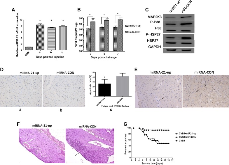
miRNA-21 overexpression inhibits CVB3 pathogenesis in mice. To detect whether high levels of miRNA-21 have effect on CVB3 infected mice, 2 × 10⁸ transduction units of lenti-miRNA-21 were intravenously injected into mice via the caudal vein. Each mice were then inoculated intraperitoneally with a dose of 1 × 10⁵ plaque forming units (PFU, LD50) of CVB3 virus (miR21-up as shown in the figure). Mice transduced with Lenti-CON followed by CVB3 infection were used as control group (miR-con as shown in the figure). A Lenti-miRNA-21 were intravenously injected into mice and miRNA-21 levels were increased significantly in the heart. B CVB3 viral titers detection were conducted at indicated time point in the hearts of mice after treatment with Lenti-miRNA-21 (n = 5), and viral titers were decreased compared with control group. C Western blot of MAP2K3, P-P38 MAPK, and P-HSP 27 expression in the heart of mice transduced with Lenti-miRNA-21. GAPDH was loaded as control group. Shown were representative results from 3 independent tests. D TUNEL assay were performed at day 7 post infection and percentage of TUNEL positive cells in each sample were shown, representative images were shown, original magnification, ×200. E Immunohistochemistry for cleaved-caspase 3 at day 7 post infection were detected, representative immunohistochemistry observation of each group was shown, examples of cleaved-caspase 3 expression are shown (black arrow), original magnification, ×200. F Histological analysis in the heart tissues at day 7 post infection of mice pretreated with Lenti-miRNA-21 or control. Examples of necrosis, calcification are shown (black arrow). Original magnification, ×200. G Evaluation of the antiviral effects of Lenti-miRNA-21 on mice survival rate. Mice were pretreated with 2 × 10⁸ transduction units of lentivirus (miR21-up and miR-con as shown in the figure) or MEM (CVB3 as shown in the figure) as indicated followed by infection of LD50 CVB3 and survival was evaluated, (*p < 0.05)

