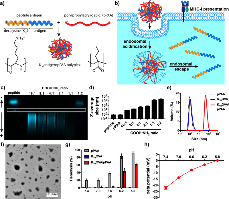
Figure 1. Fabrication and characterization of poly(propylacrylic acid)/peptide nanoplexes for enhancing MHC-I antigen presentation.
(a) Assembly of antigen-loaded nanoplexes via simple and rapid mixing of decalysine-modified antigenic peptides and pPAA, which generates electrostatically-stabilized nanoparticles. (b) Schematic representation of nanoplexes promoting cytosolic antigen delivery via endosomal escape, resulting in enhanced levels of antigen presentation on class I major histocompatibility complex (MHC-I). (c) Horizontal, native PAGE of TAMRA-labeled K10OVA and mixtures of K10OVA with pPAA at various COOH:NH2 ratios. White box outlines lanes for gel loading. (d) Z-average diameter of indicated formulations as measured by dynamic light scattering (DLS). (e) Representative size distribution (volume average) measured by DLS of soluble pPAA, soluble K10OVA, or K10OVA/pPAA polyplex generated at 2:1 COOH:NH2. (f) Transmission electron micrograph of K10OVA/pPAA polyplex generated at 2:1 COOH:NH2. (g) Erythrocyte lysis assay demonstrating pH-dependent membrane destabilizing activity of pPAA and K10OVA/pPAA polyplex. (h) ξ-potential of K10OVA/pPAA polyplexes as a function of pH.

