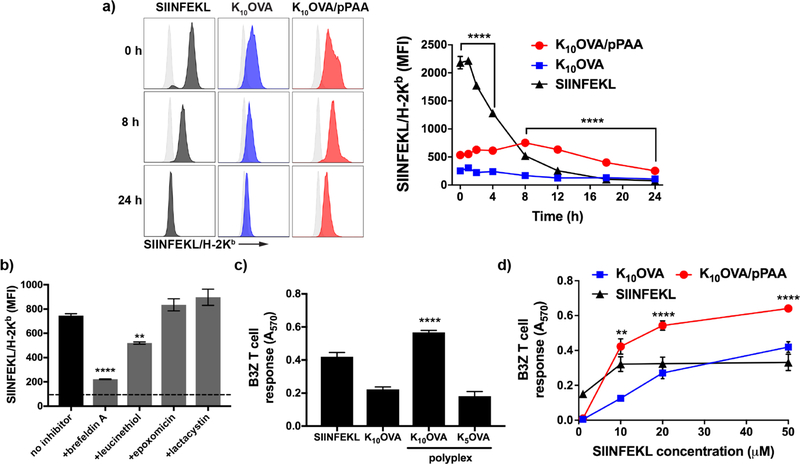
Figure 3. pPAA/peptide nanoplexes enhance and prolong antigen presentation on class I MHC.
(a) Representative flow cytometry histograms (left) and MFI over time of DC2.4 cells treated for 4 h with K10OVA, K10OVA/pPAA polyplexes, or the exact class I epitope SIINFEKL at 10 μM peptide, followed by washing, culture for time indicated, and staining with an antibody (25-D1.16) specific to the SIINFEKL/H-2Kb complex. ****p<0.0001 by one-way ANOVA with Tukey post-hoc analysis for multiple comparison. Statistical comparison between K10OVA/pPAA and SIINFEKL are shown; K10OVA/pPAA is significantly lower than SIINFEKL between 0 and 4 h and significantly higher between 8 and 24h. (b) Thirty minutes prior to 4 h treatment with K10OVA/pPAA polyplexes at 10 μM peptide, DC2.4 cells were treated with the indicated inhibitor of antigen processing or presentation followed by staining with 25-D1.16 and quantification of relative levels of SIINFEKL presentation by flow cytometry. Treatment with brefeldin A and leucinethiol inhibited antigen presentation. Dashed line represents baseline MFI of untreated DC2.4 cell; ****p<0.001, **p<0.01 relative to no inhibitor group. (c) DC2.4 were treated with the indicated formulation for 4 h at 10 μM peptide and subsequently co-cultured for 24 h with B3Z hybridoma T cells. ****p<0.001 relative to all other groups by one-way ANOVA with Tukey post-hoc test. (d) B3Z T cell response to DC2.4 cells treated with indicated formulation at different peptide concentrations. Statistical significance between K10OVA/pPAA and SIINFEKL are shown; **p<0.01, ****p<0.001 by one-way ANOVA with Tukey post-hoc test.

