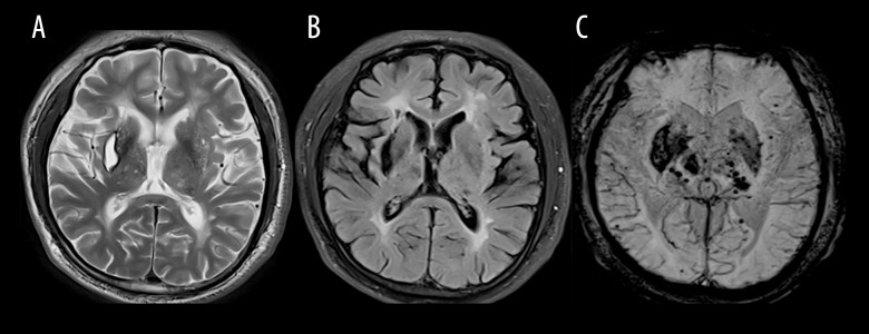
Figure 4.
(A) T2-weighted magnetic resonance (MR) image showing an old hemorrhage in the right external capsule in index patient 5; (B) FLAIR sequence showing severe leukoencephalopathy in the anterior and posterior horn of lateral ventricle and an old hemorrhage in the right external capsule; (C) Susceptibility-weighted imaging (SWI) showing an old hemorrhage in the right external capsule and previous hemorrhage in the thalamus-capsular area and numerous microbleeds (MBs) in the basal ganglia.

