
Figure 2.
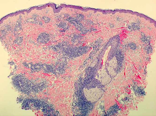
Histology of Lupus erythematosus tumidus: superficial and deep perivascular and periadnexal lymphocytic infiltrates with prominent mucinous dispositions and without epidermal changes.
Official websites use .gov
A
.gov website belongs to an official
government organization in the United States.
Secure .gov websites use HTTPS
A lock (
) or https:// means you've safely
connected to the .gov website. Share sensitive
information only on official, secure websites.
Histology of Lupus erythematosus tumidus: superficial and deep perivascular and periadnexal lymphocytic infiltrates with prominent mucinous dispositions and without epidermal changes.