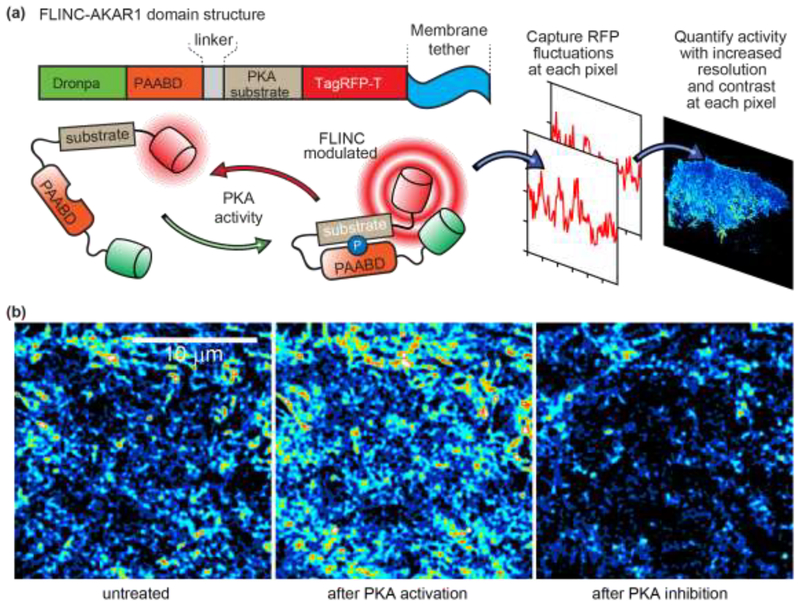
Figure 2. Working principle of the membrane-tethered FLINC–AKAR1 biosensor for photochromic Stochastic Optical Fluctuation Imaging (pcSOFI).
a) The phosphoamino-acid binding domain (PAABD) binds to its substrate upon phosphorylation by PKA, which brings TagRFP-T (red cylinder) into close proximity to Dronpa (green cylinder). The resulting changes in the fluorescence fluctuations kinetics of TagRFP-T provide a quantifiable FLINC signal of the phosphorylation state of the biosensor. b) SOFI images of FLINC–AKAR1 biosensors on the plasma membrane of live HeLa cells. After normalization to account for variability in biosensor concentration, the intensity in each pixel is the local FLINC signal, which reports on the local PKA activity. The normalized SOFI images show that PKA activity is spatially localized into distinct micro-domains (left image), and that PKA activity increases upon chemical activation (middle image) and decreases upon chemical inhibition (right image). Figure reproduced and adapted with permission from Ref. [22].

