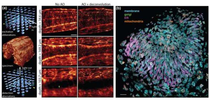
Figure 4. Adaptive optics corrected lattice-light sheet microscopy (AO-LLSM) enables live-cell imaging in whole animals.
(a) Left Panel: Imaging a 170 μm x 185 μm x 135 μm volume in the spine of a zebrafish embryo (center) at diffraction-limited resolution requires local adaptive optics correction of specimen-induced aberrations. Shown are the tiled correction wavefronts applied in the excitation (top) and emission (bottom) light paths. Larger amplitudes of the correction wavefronts are indicative of larger specimen-induced aberrations that occur at greater imaging depths. Right Panel: Slices through the image volume at different imaging depths. There is notable improvement in resolution after adaptive optics correction and deconvolution of images. Scale bar: 30 μm. b) Exploded view of hundreds of individual cells in the zebrafish eye. Four different cellular components were simultaneously labeled. The plasma membrane was labeled with a genetically-targeted citrine, the trans-Golgi apparatus was labeled with genetically-targeted mNeonGreen, the endoplasmic reticulum was labeled with genetically-targeted tagRFPt, and mitochondria were labeled with the organelle specific dye MitoTracker Deep Red. Scale bar: 30 μm. Figures reproduced and adapted with permission from Ref. [49].

