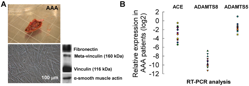
Figure 1.
(A) A sample AAA tissue (n = 20 patients) used for SMC cultures. Representative phase-contrast microscopy of SMCs obtained by explant from human AAA biopsies. Western blot analysis of SMC for fibronectin, meta-vinculin (160 kDa), vinculin (116 kDa) and α-smooth muscle actin (α-SMA). Scale bar: 100 μm. (B) mRNA expression ratio of genes (2−dCt; normalized to house-keeping genes) in SMCs derived from AAA (n = 12) patients, potentially involved in matrix degrading processes in AAA, as determined by RT-PCR.

