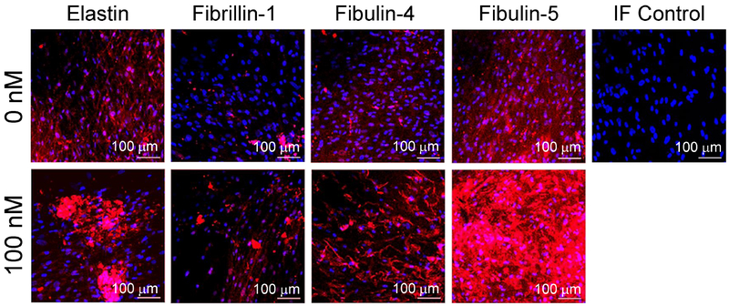
Figure 5.
Representative immunofluorescence images of AAA-SMC layers in 2D cultures, with or without GSNO, stained with respective primary antibodies for elastin, fibrillin-1, fibulin-4 and fibulin-5, and counterstained with DAPI for cell nuclei. A significant increase in the expression of all the proteins was noted in the presence of 100 nM GSNO. Scale bar: 100 μm. For each treatment condition, at least three independent cell layers were imaged, with six random regions captured in each cell layer.

