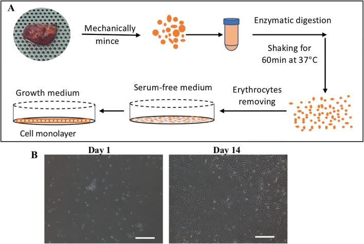
Fig. 1.
Isolation of primary human thyroid follicular epithelial cells. A Schematic isolation protocol. Non-tumorous thyroid tissues were minced mechanically into small pieces, digested with collagenase, and plated into medium without FBS for 3 days to minimize the contamination by fibroblasts. B The cell morphologies of primary isolated cells on days 1 and 14. The primary isolated cells grew as monolayers after the addition of medium with FBS. Scale bar = 500 µm

