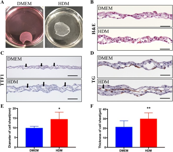
Fig. 3.
Fabrication of a thyroid cell sheet. A Cell sheets cultured on TRCDs in DMEM and HDM. B H&E staining of sheet cross-sections. C, D Immunostaining of TTF1 and TG of the sheet cross-sections. The black arrows show the follicle structure of the cell sheet. Scale bar = 50 lm. E Diameter of the cell sheets in DMEM and HDM (n = 5 in 3 cases). F Thickness of cell sheet cross-sections (n = 12 in 3 cases). Data are presented as the mean ± SD. *p < 0.05, **p < 0.01 (two-way t test)

