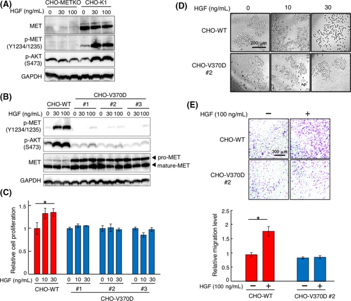
Figure 2.

Hepatocyte growth factor (HGF)‐induced responses in CHO‐WT and CHO‐V370D cells. A, Activation of MET and AKT in CHO‐K1 and CHO‐K1 MET knockout (CHO‐METKO) cells. Cells were stimulated with HGF for 10 min. MET, p‐MET, p‐AKT, and GAPDH were detected by western blotting. B, Expression and activation of MET‐WT and MET‐V370D in CHO‐METKO cells and response to HGF. Cells were stimulated with HGF for 10 min. MET, p‐MET, p‐AKT, and GAPDH were detected by western blotting. C, Cell proliferation induced by HGF. Cells were cultured with or without HGF for 48 h. Each value indicates the mean ± SE of triplicate measurements. Asterisk indicates P < .05. D, HGF‐induced cell scattering. Cells were cultured with or without HGF for 48 hours, representative images of which are shown. E, HGF‐induced cell migration. Cells were seeded on Transwell membranes and cultured for 24 h. Each value indicates the mean ± SE of triplicate measurements. Asterisk indicates P < .05. Representative images are shown
