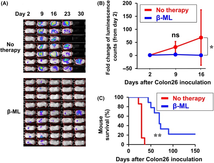
Figure 2.

Therapeutic effect of β‐ML against peritoneally disseminated colon cancer. Colon26/Luc cells were injected ip. The mice were divided into no therapy (n = 7) and β‐ML therapy (n = 9) groups. The mice in the β‐ML therapy group were injected ip with β‐ML (3 × 10⁶) from d 2, twice each week for 4 wk. A, Luminescence images are shown. B, Cancer growth of the two groups is indicated as the fold change of luminescence counts in comparison with counts observed on d 2. The linear mixed model was performed to compare the data of the no therapy and β‐ML groups, and the difference between the two groups is statistically significant (*P < .05). C, Kaplan‐Meier survival curves for β‐ML‐treated and no therapy groups are shown. The difference between the two groups is statistically significant (**P < .01, log‐rank test)
