Figure 4.

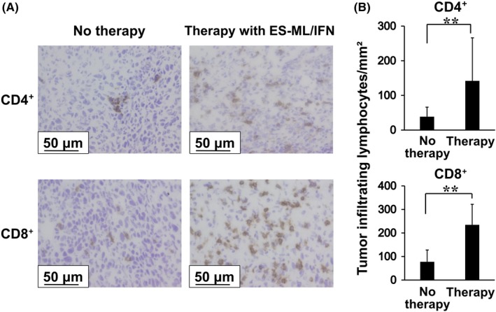
Enhanced infiltration of CD4+ cells and CD8+ cells into cancer tissues in mice treated with the ES‐ML/IFN. Colon26 cells (4 × 10⁶/mouse) were injected ip into BALB/c mice. Mice were treated from d 4, twice each week for 2 wk with both β‐ML (3 × 10⁶) and γ‐ML (7.5 × 10⁵) (n = 9) or untreated (n = 9). Then, peritoneal cancer tissues were resected and subjected to immunohistochemical analyses. A, Tissue sections were stained with anti‐CD4 and anti‐CD8 antibodies, and reactions were visualized with DAB. Scale bars represent 50 μm. B, The numbers of CD4+ cells and CD8+ cells in the cancer tissues were significantly increased in the treated mice in comparison with mice without treatment. The difference between values from the no therapy and therapy groups was evaluated using Student's t test (**P < .01)
