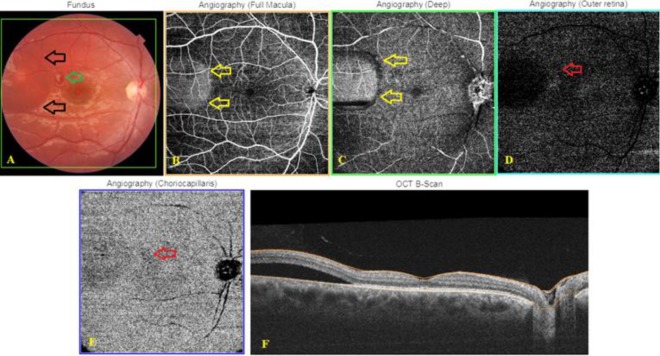
Figure 3.
Central serous chorioretinopathy in a 24-year-old man with Behçet’s uveitis while receiving systemic steroid treatment. The right eye; Color fundus picture (A), subretinal fluid at the temporal macula (black arrows) and Behçet infiltrate (green arrow). Optical Coherence Tomography Angiography (OCTA) images obtained with the Triton™ DRI swept‐source optical coherent tomography (SS‐OCT) instrument (B, C), round-well-demarcated hyperreflective area with a dark rim temporal to the fovea (yellow arrows) and minute hypoperfusion or shadowing artifact (red arrow) corresponding to the Behçet infiltrate at the level of outer retina (D) and choriocapillaris (CC) layers (E). OCT B-scan (F) demonstrating extrafoveal subretinal fluid collection

