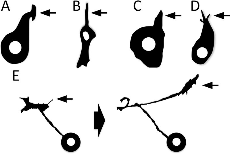
Figure 3. Development and morphology of membranous process extension and navigation.

(A–D) The extension of cellular processes (arrows) – from neutrophil (A) and mesenchymal (B) cells in vitro to bristle (C) and neuronal (D) cells in vivo. Adapted from [74,104–106]. (E) Note also the means by which axons extend “new” processes/change the direction of their process extension – via a new protrusion/filopodium (E, left arrow), which then forms the new growing cellular process/axon (E, right arrow). Adapted from [87].
