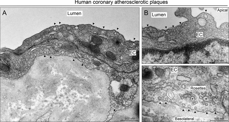
Figure 2. Caveolae morphology and localization in the endothelium of human atherosclerotic coronary arteries.
Representative EM image showing different caveolae localization at apical (A, B), intracellular (A) and basolateral side (A, C) of arterial ECs. Arrows indicate classical caveolae localized in apical and basolateral sites of ECs. Stars indicate intracellular fusion of caveolae known as “caveolae rosettes”.

