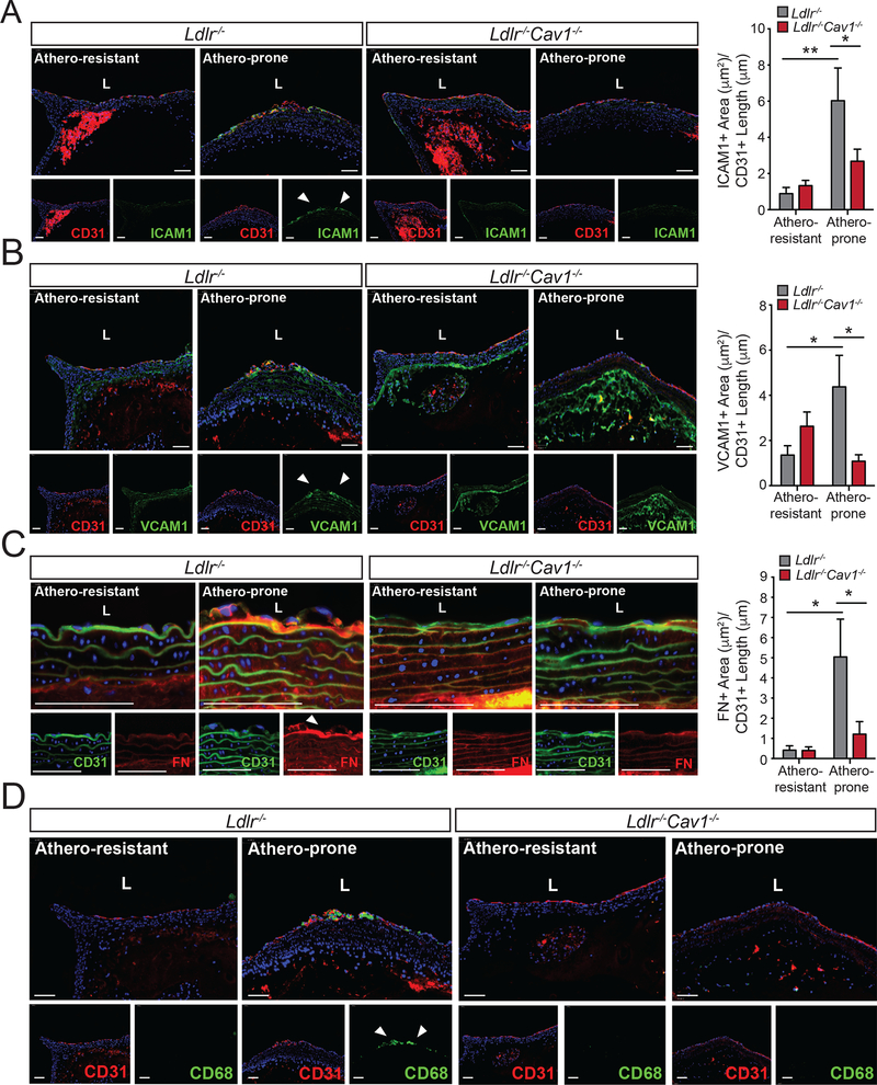
Figure 6. Absence of Cav1 attenuates vascular inflammation and macrophage infiltration in athero-prone regions of the aortic arch.
(A-C) Representative immunofluorescence analysis of athero-prone and athero-resistant areas from of 2 months old Ldlr−/− and Ldlr−/−Cav1−/− mice stained with ICAM1 and CD31 (A), VCAM1 and CD31 (B) and FN and CD31 (C). Quantifications are shown in right panels and represent the mean ± SEM (n=5–7 mice per group) of ICAM1, VCAM1 and FN positive area normalize per CD31+ length. (D) Representative immunofluorescence images of CD68 staining in the athero-prone areas of 2 months old Ldlr−/− and Ldlr−/−Cav1−/− mice fed a WD for 3 weeks. Data presented in A, B and C were analyzed by one-way ANOVA with Bonferroni correction for multiple comparisons. *P<0.05 and **P<0.01. Scale bar: 100 μm.

