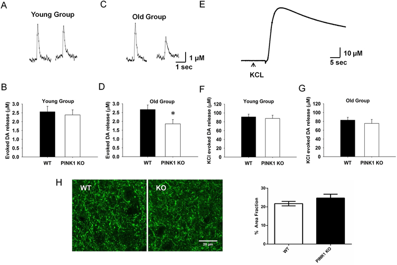
Fig. 1. Age-dependent decrease of evoked DA release in PINK1 KO dSTR slices.
Representative traces for 1p-evoked DA release in dSTR slices from both WT and PINK1 KO mice using FSCV were showed for young group (A) and old group (C) respectively. No significant difference of 1p-evoked DA release was found between PINK1 KO and WT in the young group (B, N = 6, n = 15), whereas it was significantly decreased (~30 %) in PINK1 KO in the old group (D, N = 7, n = 22). (E) Representative trace for KCl-evoked total DA release, no significant KCl-evoked DA overflow was found in either young (F, N = 5, n = 19) or old group (G, N = 4, n = 19). (H) No degeneration of DA axon terminals in PINK1 KO mice in the old group. Left panel, representative images of DA axon terminals in the striatum from WT and KO mice labeled by TH; right panel, quantification of TH-labeled DA terminals as fraction of the striatum area (N = 3 for each genotype). * p < 0.05.

