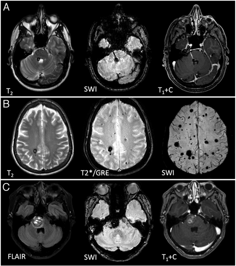
FIG. 2.
MRI features of CA. A: MRI features of a solitary CA at the floor of the 4th ventricle, clustered around a developmental venous anomaly traversing the pons. B: Autosomal dominant familial multifocal CAs, including punctate lesions on SWI, which are not seen on conventional (T2 and GRE-weighted) sequences. C: Pontine CA with the characteristic features of symptomatic hemorrhage, with acute blood expanding the lesion with a surrounding fluid-attenuated inversion recovery (FLAIR) signal of edema (left). T1+C = contrast-enhanced T1-weighted image.

