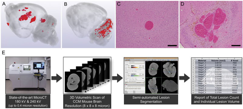
FIG. 5.
A: 3D volumetric micro-CT scan of a mouse brain with a rich repertoire of CA lesions, which developed by the 3rd month of life in the Ccm3± Trp53−/− model. B: 3D volumetric micro-CT of a mouse brain with a robust cluster of CA lesions, which developed in the hindbrain by the 10th day of life, after tamoxifen injection on postnatal day 1, inducing an endothelial Ccm3−/− state in mice expressing endothelial-specific Pdgfb promoter–driven tamoxifen-regulated Cre recombinase in combination with loxP-flanked Pdcd10 exon 4. C: Photomicrograph illustrating the histological characteristics of a primordial CA lesion, consisting of a single ballooned capillary, without bleeding, a nonheme iron deposit, or inflammatory cell infiltrate (H&E, bar = 200 μM). D: Multicavernous mature CA with all the histological features of human lesions with hemosiderin deposits (H&E, bar = 200 μM). E: Process of image acquisition by micro-CT, 3D reconstruction, and semiautomated volumetric assessment of the lesion burden.

