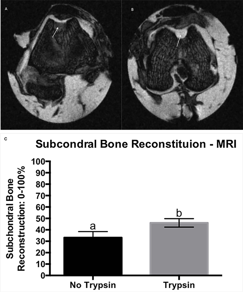
Figure 1.

Trasnverse PDFS MRI image showing improved subchondral bone reconstitution (arrows) of the reparative tissue treated with trypsin (B), compared to its contralateral control (A), MRI score for main effect of trysin on subchondral bone reconstitution, grading scale 0–100%, where 0% is no subchrondral bone reconstitution and 100% is subchondral bone reconstituted to the level of surrounding tissue. Error bar represents standard error. Different letters mean statistical siginificant difference (C).
