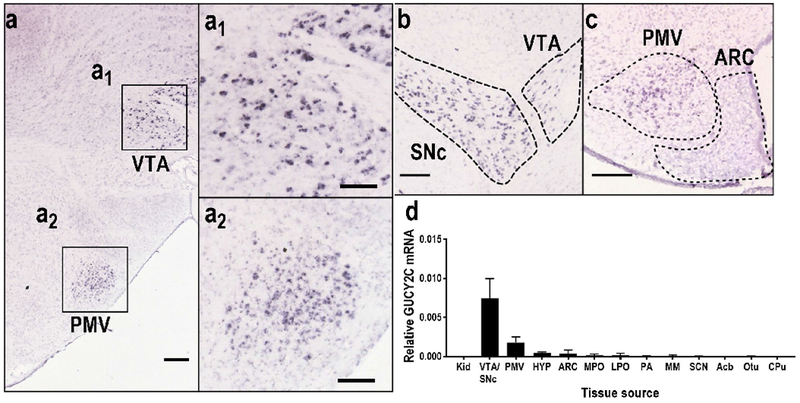
CNS GUCY2C mRNA expression is restricted to the hypothalamic ventral premammillary nucleus and neurons in the VTA/SNc. (a, sagittal; b-c, coronal)
In situ hybridization (Allen Institute for Brain Science; experiments 73992911 and 69734851) (
Lein et al. 2007) revealed robust GUCY2C mRNA expression in the PMV and the VTA/SNc, while mRNA was absent in the ARC. (d) Quantitative RT-PCR of microdissected hypothalamic nuclei or tissue controls demonstrated that hypothalamic GUCY2C mRNA is restricted to the premammillary nucleus. For each region, n=5 mice per group, with mRNA analyzed in duplicate; data represent mean +/− SD. VTA/SNc: Ventral Tegmental Area/Substantia Nigra, pars compacta; PMV: Ventral Premammillary nucleus; hyp: bulk hypothalamus; ARC: Arcuate nucleus; MPO: Medial Preoptic nucleus; LPO: Lateral Preoptic nucleus; PA: Posterior Amygdalar nucleus; MM: Medial Mamillary nucleus, SCN: Suprachiasmatic nucleus; Acb: Nucleus Accumbens; OTu: Olfactory Tubercle; Cpu: Caudoputamen. Scale bars = 200 μm in a-c; 100 μm in al and a2. Source images for (a), (b), and (c) can be found at the following links:

