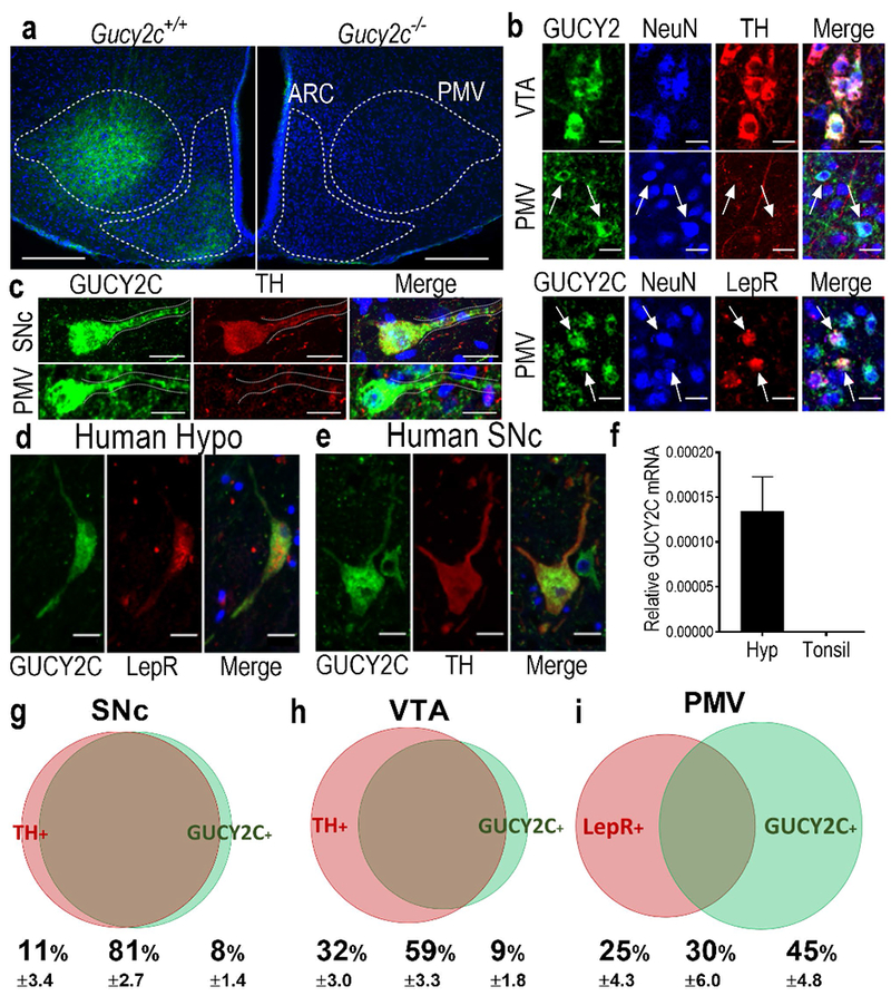
Figure 2.

Hypothalamic neurons in the PMV express GUCY2C protein in somas and projections. (a) Immunofluorescence of mouse hypothalamus revealed a subpopulation of GUCY2C (green)-expressing cells emanating from the PMV, with expression in adjacent arcuate nucleus, in Gucy2+/+, but not Gucy2c−/− mice. Blue: DAPI. (b) GUCY2C(+) cells in the VTA and PMV express the neuronal marker NeuN. While GUCY2C(+) neurons in the VTA express TH by immunofluorescence, GUCY2C(+) neurons in the PMV are TH(−), and may be LepR(+). Arrows indicate PMV NeuN(+), GUCY2C(+) cells that do not express TH (middle column), or express LepR (bottom column). (c) Neurons in the SNc (top) and PMV (bottom) express GUCY2C protein in cell bodies and neuronal processes (dotted line). GUCY2C expression in human hypothalamus and midbrain matches the pattern of expression in mice. (d) In humans, hypothalamic GUCY2C+ neurons are LepR(+) while (e) midbrain GUCY2C neurons are TH(+). (f) GUCY2C mRNA is expressed in human hypothalamus, but not tonsil (negative control); n=4 human hypothalamic samples (see Suppl. Table 1 for details); for tonsil samples, n=3. Neuronal enumeration revealed that in the: (g) SNc, 88% of TH(+) neurons (red bubble) are GUCY2C(+) (green bubble); (h) VTA, 65% of TH(+) neurons are GUCY2C(+); and (i) PMV, 46% of LepR(+) neurons (red bubble) are also GUCY2C(+) (green bubble). Bubble size and overlap are relative to the combined total of GUCY2C(+) and TH(+) or LepR(+) neurons in the VTA/SNc or PMV, respectively. Scale bars: a: 200 μm; b-f: 20 μm; F inset: 5 μm.
