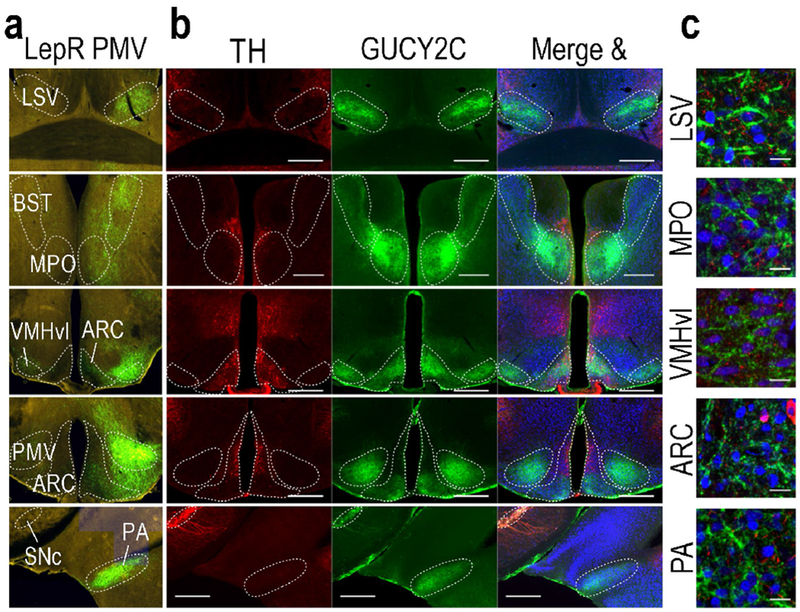
Figure 3.

PMV LepR projection sites express GUCY2C protein in projections. (a) Unilateral stereotaxic injection of cre-inducible GFP AAV into the PMV of LepR-cre mice reveals GFP(+) projections in hypothalamic and extrahypothalamic nuclei (Allen Institute for Brain Science, experiment ID 167656388)(Oh et al. 2014). (b) GUCY2C is expressed in PMV projection sites of LepR neurons, but not with TH. (c) High magnification reveals that GUCY2C protein is expressed in projections, not neuronal cell bodies in hypothalamic nuclei other than the PMV Scale bars in (b): 200 μm; in C: 20 μm. LSV: lateral septal nucleus, ventral part; BST: bed nucleus of the stria terminalis; MPO: medial preoptic nucleus; vl: ventrolateral aspect of the ventromedial hypothalamus; ARC: Arcuate nucleus; SNc: substantia nigra, pars compacta; PA: posterior amygdalar nucleus. Source images for panel (a) can be found at the following links:
