
Figure 5.
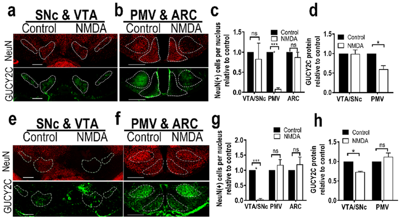
Selective stereotaxic ablation of the PMV or VTA/SNc with NMDA reduced GUCY2C-immunofluorescent neurons specifically in the targeted region. (a) Representative unilateral ablation of PMV with NMDA did not affect neurons or GUCY2C immunofluorescence in the SNc or VTA. (b) In contrast to the control side (left), NMDA injection eliminated neurons in the PMV, but not in the ARC. (c) Stereotaxic PMV ablation with NMDA significantly reduced the number of neurons [NeuN(+) cells] in the PMV, without reducing the number of neurons in the SN/VTA or arcuate nucleus, (d) Quantification of GUCY2C protein expression in nuclei expressing GUCY2C(+) cell bodies revealed that unilateral PMV ablation eliminated GUCY2C protein expression in the PMV, but not in the VTA or SNc; n=2 mice, (e) Unilateral injection of NMDA into the VTA/SNc severely diminished GUCY2C-expressing neurons in the VTA/SNc while sparing VTA/SNc neurons on the control side, (f) NMDA injection into the VTA/SNc did not affect neurons in the PMV or ARC. (g) Stereotaxic VTA/SNc ablation with NMDA significantly reduced the number of neurons in the VTA/SNc, but not the PMV or ARC. (h) Quantification of GUCY2C protein expression in nuclei expressing GUCY2C(+) cell bodies revealed that unilateral VTA/SNc ablation eliminated GUCY2C expression in the VTA/SNc, but not the PMV or ARC. Scale bars in (a), (b), (e), and (f): 200 μm; *p<0.05; ****p<0.0001.