Figure 6.

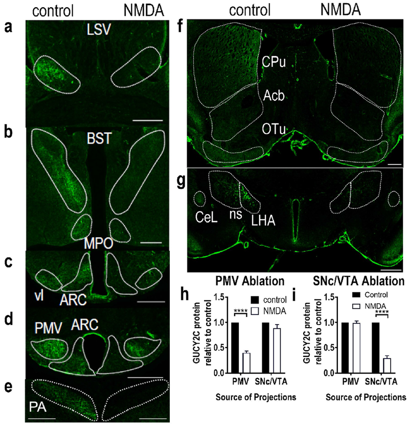
Selective stereotaxic ablation of the PMV or VTA/SNc with NMDA eliminated GUCY2C immunofluorescence specifically in distant projections from the targeted region. NMDA ablation of the PMV eliminated GUCY2C immunofluorescence in projections from the PMV: (a) LSV, (b) BST and MPO, (c) vl and rostral ARC, (d) PMV and caudal ARC, and (e) PA. Conversely, NMDA ablation of the VTA eliminated GUCY2C immunofluorescence in projections from the VTA: (f) CPu, Acb, and OT, and (g) LHA, ns, and CeL. NMDA ablation of the PMV (h) or VTA/SNc (i) produced selective loss of GUCY2C protein expression in regions canonically associated with projections from these nuclei, respectively. Data in (h) and (i) represent GUCY2C protein expression normalized to the contralateral control side. For (h), data were collected from 2 mice with PMV ablation in the absence of neuronal loss in the arcuate nucleus, SNc, or VTA. For (i), data were collected from 2 mice with VTA/SNc ablation in the absence of neuronal loss in the PMV or arcuate nucleus. LSV: Lateral Septal nucleus, Ventral part; BST: Bed nucleus of the Stria Terminalis; MPO: Medial Preoptic nucleus; vl: ventromedial hypothalamus, ventrolateral part; ARC: Arcuate nucleus; PMV: Ventral Premammillary nucleus; PA: Posterior Amygdalar nucleus CPu: Caudoputamen; Acb: nucleus Accumbens; OT: Olfactory Tubercle; ns: nigrostriatal tract; LHA: Lateral Hypothalamic Area; CeL: Lateral part of the Central amygdalar nucleus. Scale bars in (a)-(g): 200 μm. ****p<0.0001.
